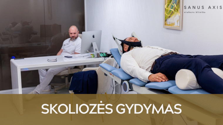
Sanus Axis Clinic treats scoliosis using individually tailored spinal correction and muscle balance restoration programs.
Our specialists assess the degree and structure of the spinal curvature, as well as the biomechanical factors contributing to it, in order to determine the underlying cause of the deformity and evaluate the risk of progression.
Why Patients Choose Sanus Axis:
- The clinic employs highly qualified specialists in manual therapy and physiotherapy;
- We use advanced medical equipment unavailable elsewhere in Lithuania;
- Treatment combines manual therapy, spinal decompression, physiotherapy, and neural mobilization techniques to restore natural spinal biomechanics;
- The average treatment course consists of only five sessions, after which patients are able to return to their normal daily activities.
What Is Scoliosis?
Scoliosis is a sideways curvature of the spine in which the vertebral column develops a C-shaped or S-shaped curve.
This deformity alters spinal biomechanics, causes muscle imbalance, postural asymmetry, and back pain, and in more severe cases may also affect respiratory function.
Scoliosis is most commonly diagnosed during childhood or adolescence, but it can also develop in adults due to degenerative changes, physical trauma, or long-term muscle imbalance.
Early treatment helps control curve progression and reduces both functional and cosmetic complications.
Symptoms of Scoliosis
Scoliosis most commonly presents with visible body asymmetry and postural changes, including:
- One shoulder positioned higher than the other;
- One shoulder blade protruding more prominently;
- A deeper waist crease on one side of the body;
- Pelvic tilt or pelvic rotation;
- A visibly curved spinal line when viewed from behind;
- A noticeable lean of the torso to one side while standing or walking;
- A rib hump or muscular prominence in the lumbar region when bending forward;
- Back fatigue or pain after prolonged sitting or standing;
- Clothing fitting unevenly, such as asymmetrical shoulder alignment.
Degrees of Scoliosis According to the Cobb Angle
The Cobb angle is the standard method used to measure the degree of spinal curvature in scoliosis.
It is assessed using an X-ray by drawing lines along the most tilted vertebrae and calculating the angle formed at their intersection.
This measurement helps determine whether scoliosis is mild, moderate, or severe, assess the risk of progression, and select the most appropriate treatment plan.
| Degree (Cobb Angle) | Description | Functional / Visual Signs | Treatment Approach |
|---|---|---|---|
| <10° – Postural asymmetry | Slight deviation of the spinal line with minimal vertebral rotation. This is not yet considered scoliosis. | Mild differences in shoulder or pelvic height; asymmetry visible only in certain positions | Monitoring, patient education on proper posture, and preventive physiotherapy. |
| 10–25° – Mild scoliosis | Clearly visible lateral curvature with mild vertebral rotation. | Shoulder, waist, or shoulder blade asymmetry; postural changes may progress during growth in children and adolescents. | Individualized physiotherapy, asymmetric corrective exercises combined with breathing techniques, and regular monitoring. |
| 25–40° – Moderate scoliosis | More pronounced deformity with a high risk of progression during growth spurts. | Visible rib hump, pelvic rotation, trunk asymmetry, fatigue, or muscle tension. | Intensive physiotherapy based on an individualized treatment plan; bracing is often recommended. |
| >40° – Severe scoliosis | Significant lateral curvature with marked vertebral rotation and possible effects on internal organ function. | Severe trunk deformity, chest wall changes, and restricted breathing. | Surgical evaluation is recommended to determine whether corrective surgery is necessary If surgical intervention is not clearly indicated or surgery is not yet required, specialized physiotherapy is typically recommended. |
Types of Scoliosis
Idiopathic Scoliosis
Idiopathic scoliosis is the most common form of scoliosis, accounting for approximately 80–90% of all cases
Its exact cause remains unknown, although research suggests a strong genetic component, which is why the condition often occurs across multiple generations within the same family. Idiopathic scoliosis is most commonly diagnosed during childhood or adolescence, particularly during periods of rapid spinal growth, when the risk of progression is highest.
This type of scoliosis usually does not cause pain initially and is most often first noticed because of body asymmetry. When detected early, idiopathic scoliosis can be effectively managed through physiotherapy, corrective exercises, and other non-invasive treatment methods.
Degenerative Scoliosis
Degenerative scoliosis develops in adulthood as a result of age-related changes in spinal structures.
As the body ages, intervertebral discs lose height, facet joints degenerate, and ligaments become less elastic. These changes reduce spinal stability and may lead to lateral spinal curvature. Degenerative scoliosis is most commonly diagnosed in individuals aged 50–60 years, particularly in postmenopausal women.
This form is often associated with back pain, gait disturbances, numbness or weakness in the legs due to nerve irritation. Treatment focuses on pain relief, restoring function, and controlling progression through manual therapy, neural mobilization, and specialized physiotherapy techniques.
Congenital Scoliosis
Congenital scoliosis occurs when the vertebrae do not form properly during fetal development.
This may involve fused vertebrae, wedge-shaped vertebrae, or other structural abnormalities that cause spinal deformity from early childhood. Congenital scoliosis often progresses more rapidly than other forms because the deformity worsens as the child grows.
This condition requires ongoing orthopedic supervision, detailed diagnostic evaluation, and individually tailored treatment to reduce progression and prevent functional spinal complications.
Neuromuscular Scoliosis
Neuromuscular scoliosis develops as a result of neurological or muscular disorders that impair the function of the muscles responsible for spinal stability.
It is most commonly associated with conditions such as cerebral palsy, muscular dystrophy, spinal cord injuries, or other neurological disorders. Due to impaired muscle tone and control, the spinal curvature may progress rapidly and can become severe in both the thoracic and lumbar regions.
The primary goals of treatment are to improve trunk stability, respiratory function, and overall quality of life. Therapy is typically combined with comprehensive rehabilitation programs.
Functional Scoliosis
Functional scoliosis develops not because of structural abnormalities in the vertebrae, but due to external factors that temporarily alter spinal alignment.
Common causes include leg length discrepancy, muscle imbalance, pelvic rotation, postural disorders, or compensation for pain. Functional scoliosis is not fixed — once the underlying cause is addressed and appropriate physiotherapy is applied, the spine can return to a neutral position.
This is the most treatable form of scoliosis, as early intervention can fully restore normal spinal biomechanics and prevent progression of the deformity.
Scoliosis Diagnosis
At Sanus Axis Clinic, scoliosis diagnosis is performed using a comprehensive approach that evaluates not only X-ray findings but also functional parameters that help identify the true cause of the deformity and assess the risk of progression.
The Diagnostic Process Includes:
- X-ray evaluation (Cobb angle measurement) – used to determine the degree of spinal curvature, vertebral rotation, and the nature of the deformity;
- MRI (when indicated) – performed when congenital vertebral abnormalities, nerve structure involvement, or atypical curve progression are suspected;
- Biomechanical spinal assessment – analysis of posture, pelvic alignment, spinal mobility, muscle imbalance, and load distribution contributing to scoliosis progression;
- Functional and neurological testing – assessment of muscle strength, balance, trunk control, breathing mechanics, and nervous system function;
- Physiotherapy consultation – integration of all diagnostic findings to create an individualized scoliosis management and treatment plan.
Rehabilitation After Scoliosis Treatment
Rehabilitation following the initial phase of scoliosis treatment is essential to stabilize the achieved correction, restore symmetrical muscle function, and reduce the risk of further curve progression.
Goals of Rehabilitation
- Stabilize the spine after treatment and maintain the achieved correction;
- Strengthen the deep core muscles responsible for supporting symmetrical posture;
- Improve chest mobility and breathing mechanics, particularly in cases involving a rib hump;
- Reduce residual muscle tension and compensatory movement patterns;
- Integrate corrective strategies into daily activities such as sitting, walking, exercising, and lifting.
Rehabilitation Process
- Stabilization Phase – corrective breathing exercises and isometric exercises are performed to help maintain spinal alignment without additional external support.
- Functional Strengthening Phase – patients learn how to maintain proper spinal alignment during everyday activities, including sitting, standing, lifting objects, and participating in sports.
- Integration Phase – patients learn how to maintain proper spinal alignment during everyday activities, including sitting, standing, lifting objects, and participating in sports.
- Maintenance Phase - the physiotherapist develops a long-term home exercise program aimed at preserving spinal correction and muscular symmetry, while also establishing a schedule for periodic follow-up assessments to prevent curve progression.
Rehabilitation Duration
The duration of rehabilitation depends on the degree of scoliosis, the patient’s age, and the initial progression of the spinal curve.
In most cases, the maintenance program continues for approximately 2 to 12 months, with exercises periodically adjusted according to the patient’s progress.
When Is It Necessary to Seek Medical Attention?
- You notice that one shoulder or shoulder blade is significantly higher than the other;
- A child or adolescent develops noticeable postural changes, especially during periods of rapid growth;
- A rib hump or pronounced waist asymmetry becomes visible when bending forward;
- Back pain develops and worsens after prolonged sitting or standing;
- Child complains of fatigue, balance difficulties, or discomfort after physical activity;
- You notice asymmetry in the way clothing fits (e.g., one shoulder strap constantly slips down, shirts twist to one side);
- Scoliosis has already been diagnosed, but there are signs that the spinal curve may be progressing.
Scoliosis Prevention
- Monitor posture. Keep the shoulders level, avoid consistently leaning to one side, and do not carry a backpack on one shoulder only;
- Ensure an ergonomic work and study environment. The monitor should be at eye level, the chair adjusted to the person’s height, and the feet placed flat on the floor; for children, proper desk-to-chair proportions are especially important;
- Move regularly. Daily walking, swimming, or physiotherapy exercises help maintain muscle symmetry and reduce the risk of overload;
- Strengthen the core muscles. Balanced back and abdominal muscles help maintain proper spinal alignment and compensate for asymmetries;
- Avoid prolonged static positions. Sitting or standing in the same position for extended periods increases muscle imbalance — take breaks every 45–60 minutes;
- Monitor periods of rapid growth in children. During growth spurts, the risk of scoliosis progression increases, so parents should regularly assess the symmetry of the shoulders, shoulder blades, waist, and pelvis.
Frequently Asked Questions
Can scoliosis progression be stopped without a brace?
Yes — the progression of mild to moderate scoliosis can often be controlled through specialized physiotherapy, corrective exercises, and ergonomic adjustments in daily activities. A brace is only necessary when the Cobb angle and growth rate indicate a high risk of progression.
Can scoliosis cause internal organ dysfunction?
Yes — spinal curves greater than 40° may reduce chest cavity volume, impair breathing, affect cardiac function, and limit physical capacity. Early treatment helps prevent these complications.
Is scoliosis always visible to the naked eye?
Not always. Early-stage scoliosis is often identified through postural assessment, X-rays, and biomechanical evaluation. During periods of rapid growth, parents should monitor the symmetry of the shoulders, shoulder blades, and waist, and consult a specialist if any changes are noticed.
Can people with scoliosis participate in sports?
Yes. People with scoliosis can participate in sports as long as the movements do not cause pain or neurological symptoms. However, sports that consistently overload one side of the body (such as tennis, badminton, or throwing sports) should be approached with caution.
When does scoliosis require surgery?
Surgery is considered when the spinal curvature exceeds 50° on the Cobb scale, progresses rapidly, or causes functional complications such as shortness of breath, breathing restrictions, neurological symptoms, or significant chest deformity. Surgical treatment is only recommended when conservative treatment methods can no longer prevent progression of the condition.